Here is all you needed to know about the Hip Joint.
The hip joint is one of the most important joints in the human body. It allows us to walk, run, and jump. It bears our body’s weight and the force of the strong muscles of the hip and leg. Yet the hip joint is also one of our most flexible joints and allows a greater range of motion than all other joints in the body except for the shoulder.
General Anatomy
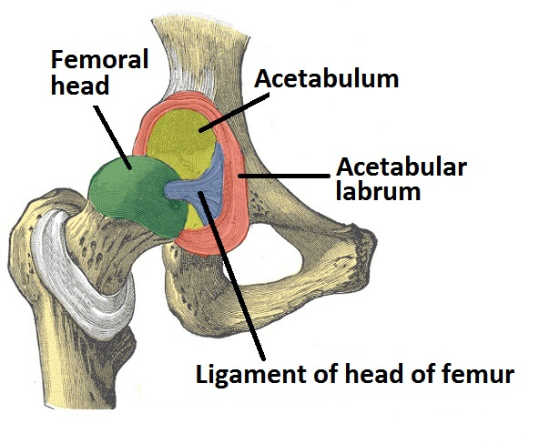
The acetabulum is a cup-like depression in the lateral side of the pelvis, much like the glenoid fossa of the scapula, only deeper. The head of femur is spherical, and hence the joint is classified as a ball and socket joint.
Both the acetabulum and head of femur are covered in articular cartilage, which is thicker at the places of weight bearing
Ligament of head of femur: A relatively small ligament that attaches from the acetabular fossa to the fovea of the femur. It encloses a branch of the oburator artery, which contributes a small proportion of the blood supply to the hip joint.
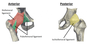
Pubofemoral: Found anteriorly and inferiorly on the hip joint. It attaches at the pelvis to the iliopubic eminance and obturator membrane, and then blends with the articular capsule. It prevents excessive abduction and medial rotation.
Iliofemoral: Found anteriorly on the hip joint. It originates from the ilium, just inferior to the anterior, inferior iliac spine. It attaches to the intertrochanteric line, thickening to two places, giving the ligament a Y shaped appearance. It prevents hyperextension of the hip joint.
Ischiofemoral: The main posterior ligament of the hip joint. It attaches to the ischium of the pelvis and the greater trochanter of the femur. It prevents excessive medial rotation of the femur at the hip joint.
Factors that Stabilise the Hip Joint
The primary function of the hip joint is weight bearing, and so there are features that increase its stability.
The first is the structure of the acetabulum. It is deep, and encompasses nearly all of the head of the femur. This decreases the probability of the head slipping out of the acetabulum, and causing a dislocation.
There is a fibrocartilaginous collar around the acetabulum which increases its depth. It is known as the acetabular labrum. The increase is depth provides a large articular surface , thus improving the stability of the joint.
The iliofemoral, pubofemoral and ischiofemoral ligaments are very strong, and along with the thickened joint capsule, they stabilise the joint greatly. These ligaments have a uniquespiral orientation; this causes them to become tighter when the joint is extended, which adds stability to the joint, and also means less energy is needed to maintain a standing position.
Muscles and ligaments work in a reciprocal fashion at the hip joint:
•Anteriorly, where the ligaments are strongest, the medial flexors (located anteriorly) are fewer and weaker.
•Posteriorly, where the ligaments are weakest, the medial rotators are greater in number and stronger – they effectively ‘pull’ the head of the femur into the acetabulum.
Movements and Muscles
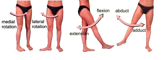
The movements that can be carried out at the hip joint are flexion, extension, abduction, adduction and medial/lateral rotation.
The degree to which flexion at the hip can occur depends on whether the knee is flexed, which relaxes the hamstrings, and increases the range of flexion.
Extension at the hip joint is limited by the joint capsule, and in particular, the iliofemoral ligament. These structures become taut during extension to limit further movement.
Listed below are the movements of the hip joint, and the principle muscles responsible for those movements:
•Flexion: Iliosoas, rectus femoris, sartorius
•Extension: Gluteus maximus, semimembranous,semitendinosus and biceps femoris
•Abduction: Gluteus medius, gluteus minimus and the deep gluteals (piriformis, gemelli etc)
•Adduction: Adductors longus, brevis and magnus,pectineus and gracillis
•Lateral rotation: Biceps femoris, gluteus maximus, and the deep gluteals (piriformis, gemelli etc)
•Medial rotation: Gluteus medius and minimus, semitendinosus and semimembranosus
Text and Images compiled by: Masoom


0 Comments